Special examinations
Laboratory studies
Patients with uncomplicated cholelithiasis or simple biliary colic typically have normal laboratory test results.
Acute cholecystitis is associated with polymorphonuclear leukocytosis.
Choledocholithiasis with acute common bile duct obstruction initially produces an acute increase in the level of liver transaminases (alanine and aspartate aminotransferases), followed within hours by a rising serum bilirubin level. If obstruction persists, a progressive decline in the level of transaminases with rising alkaline phosphatase and bilirubin levels may be noted over several days. Concurrent obstruction of the pancreatic duct by a stone in the Vater’s ampulla may be accompanied by increases in circulating lipase and amylase levels.
Repeated testing over hours to days may be useful in evaluating patients with gallstone complications. Improvement of the levels of bilirubin and liver enzymes may indicate spontaneous passage of an obstructing stone. Conversely, rising levels of bilirubin and transaminases with progression of leukocytosis in the face of antibiotic therapy may indicate ascending cholangitis.
Investigations in acute cholecystitis
Ultrasonography. Ultrasonographic findings that are suggestive of acute cholecystitis include the following: pericholecystic fluid, gallbladder wall thickening greater than 4 mm and sonographic Murphy’s sign. The presence of gallstones also helps to confirm the diagnosis (fig. 7, 8).
The sensitivity and specificity of CT/MRT scans for predicting acute cholecystitis have been reported to be greater than 95%. Spiral CT scans and MRI have the advantage of being noninvasive, but they have no therapeutic potential and are most appropriate in cases where stones are unlikely.
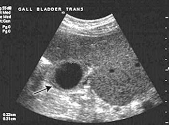
Figure 7 – Ultrasonic examination showed a thickened gallbladder wall and pericholecystic fluid
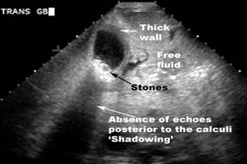
Figure 8 – Ultrasonic findings: thick gallbladder wall, tones in gallbladder, absence of echoes posterior to the calculi –“Shadowing”
Findings suggestive of cholecystitis include wall thickening (>4 mm), pericholecystic fluid, subserosal oedema, intramural gas, and sloughed mucosa (fig. 9).
Endoscopic retrograde cholangiopancreatography may be useful in patients at high risk for common duct gallstones, if signs of common bile duct obstruction are present (fig. 10).
Endoscopic retrograde cholangiopancreatography allows visualization of the anatomy and may be therapeutic by removing stones from the common bile duct. Disadvantages include the need for a skilled operator, high cost, and complications such as pancreatitis, which occurs in 3–5% of cases.
Дата добавления: 2015-07-04; просмотров: 2097;