Special examinations. Serum chemistries, liver panels
Laboratory studies
Serum chemistries, liver panels. Results are usually normal or mildly elevated.
Creatinine. Elevations may indicate dehydration.
CBC: WBC may be elevated with a left shift in simple or strangulated obstructions. Increased hematocrit speaks to volume state (i.e., dehydration).
Lactate dehydrogenase tests
Urinalysis
Imaging studies
Radiography. Order plain radiographs first for patients in whom SBO is suspected. At least 2 views, supine or flat and upright, are required. Too findings were more predictive of a higher grade or complete SBO: presence of air-fluid differential height in the same small-bowel loop and presence of a mean level width greater than 25 mm. When these findings are present, the obstruction is most likely high grade or complete. When both are absent, a low (partial)-grade SBO is likely or nonexistent. Absent or minimal colonic gas indicates SBO (fig.19).

Figure 19 – Multiple air fluid levels
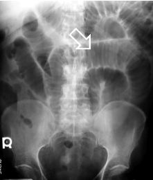
Small bowel loops contain transverse folds known as valvulae conniventes or plica circularis. These folds are well seen in this patient with small bowel obstruction. Usually the colon is decompressed and hardly visible (fig. 20).
Enteroclysis
This is valuable in detecting presence of obstruction and in differentiating partial from complete blockages. This study is useful when plain radiographic findings are normal in the

Figure 20 – Plica circularis
presence of clinical signs of SBO or if plain radiographic findings are nonspecific. It distinguishes adhesions from metastases, tumor recurrence, and radiation damage. Enteroclysis offers a high negative predictive value and can be performed with 2 types of contrast. Barium is the classic contrast agent used in this study. It is safe and useful when diagnosis obstructions provided no evidence of bowel ischaemia or perforation exists. Barium has been associated with peritonitis and should be avoided if perforation is suspected.
CT scanning
CT scanning is useful in making an early diagnosis of strangulated obstruction and in delineating the myriad other causes of acute abdominal pain, particularly when clinical and radiographic findings are inconclusive. It also has proved useful in distinguishing the aetiologies of SBO, i.e., extrinsic causes such as adhesions and hernia from intrinsic causes such as neoplasms or Crohn’s disease. It also differentiates the above from intraluminal causes such as bezoars. CT scanning is about 90% sensitive and specific in SBO diagnosis. CT scanning is the study of choice if the patient has fever, tachycardia, localized abdominal pain, and/or leukocytosis. It is capable of revealing abscess, inflammatory process, extraluminal pathology resulting in obstruction, and mesenteric ischaemia. CT scanning enables the clinician to distinguish between ileus and mechanical small bowel in postoperative patients. Obstruction is present if the small-bowel loop is greater than 2.5 cm in diameter dilated proximal to a distinct transition zone of collapsed bowel less than 1 cm in diameter (fig. 21).

Figure 21 – Note the dilated small bowel loops with a focal transition zone to distal collapsed bowel
A smooth beak indicates simple obstruction without vascular compromise; a serrated beak may indicate strangulation. Bowel wall thickening indicates early strangulation. CT scanning is useful in identifying abscesses, hernias and tumors.
Ultrasonography
Ultrasonography is less costly and less invasive than CT scanning. It may reliably exclude SBO in as many as 89% of patients. Specificity is reportedly 100%.
Ultrasonography signs of SBO:
– dilatation of small bowel lumen;
– “pendulous” movements of bowel content.
Дата добавления: 2015-07-04; просмотров: 1039;
