Special examinations. Studies are directed at evaluating the dehydration and electrolyte imbalance that may occur as a consequence of LBO and at ruling out ileus as a diagnosis
Laboratory studies
Studies are directed at evaluating the dehydration and electrolyte imbalance that may occur as a consequence of LBO and at ruling out ileus as a diagnosis. Suggestion of an abnormal anion gap also should prompt an arterial blood gas measurement and/or a serum lactate level measurement. A decreased haematocrit level, particularly with evidence of chronic iron-deficiency anemia, may suggest chronic lower gastrointestinal bleeding, particularly due to colon cancer. A stool test also should be performed, for similar reasons. Although bowel obstruction, or even constipation, may mildly elevate the WBC count, substantial leukocytosis should prompt reconsideration of the diagnosis. Ileus, secondary to an intra-abdominal or extra-abdominal infection or another process, is a possibility.
Imaging studies
Flat and upright abdominal roentgenography demonstrates dilation of the large bowel and air fluid levels. Сolonic air suggests the anatomic location of the obstruction. A dilated colon without air in the rectum is more consistent with obstruction. The presence of air in the rectum is consistent with obstipation, ileus, or partial obstruction. This finding can be misleading, particularly if the patient has undergone rectal examinations or enemas. The characteristic bird’s beak of volvulus may be seen.
If differentiation between obstipation and obstruction is required, imaging with contrast is indicated (fig. 22). If localization is required for surgical intervention, imaging with contrast is indicated. Water-soluble gastrografin has important advantages over barium as a contrast agent and generally should be employed first. It usually does not cause chemical peritonitisk, if the patient has colonic perforation.
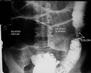
Figure 22 – Left side colonic cancer with obstruction
CT scanning is not used initially in patients with large bowel obstruction unless a diagnosis has been made. CT scan, particularly with rectal contrast, may demonstrate a mass or evidence of metastatic disease.
Other tests. Fiber-optic endoscopy may be useful in evaluating left-sided colonic obstruction, including the anatomic location and pathology of the lesion. Because the cecum is not reached in such cases, the endoscopist must be alert to the possibility of incorrectly identifying anatomic landmarks and the location of the obstruction. Although flexible endoscopy is relatively comfortable for the patient and provides a better view than rigid sigmoidoscopy, the latter also may be used, depending on the availability of resources and training of personnel. Right-sided colonic obstruction is more difficult to evaluate without first administering an oral bowel preparation, which is contraindicated in the setting of bowel obstruction.
Дата добавления: 2015-07-04; просмотров: 975;