1 страница
МЫШЕЧНАЯ СИСТЕМА
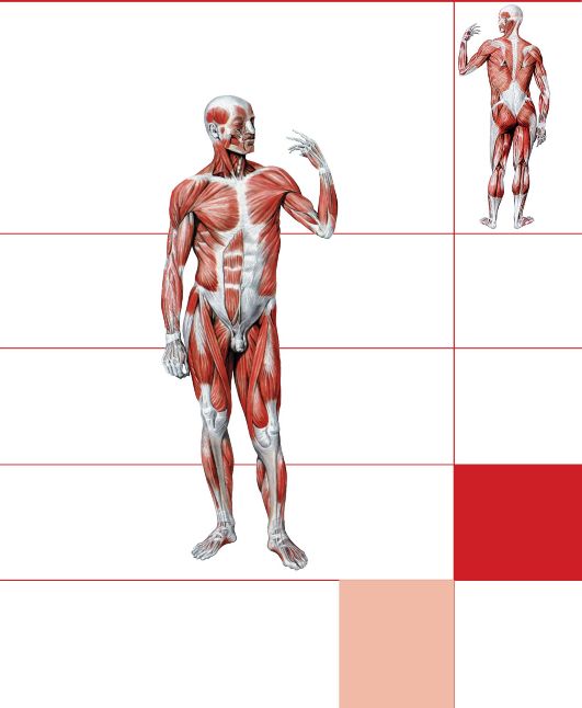
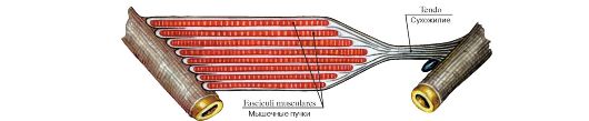 Рис. 247. Схема начала и прикрепления мышцы
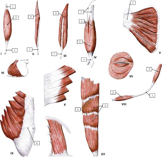 Рис. 248. Форма мышц:
I - веретенообразная мышца; II - одноперистая мышца; III - двуперистая мышца; IV - двуглавая мышца; V - многоперистая мышца; VI - треугольная мышца; VII - круговая мышца; VIII - двубрюшная мышца; IX - широкая мышца, имеющая апоневроз; X - зубчатая мышца; XI - квадратная мышца; XII - мышца, имеющая сухожильные перемычки; 1 - сухожилие; 2 - брюшко; 3 - головка; 4 - апоневроз; 5 - сухожильная перемычка
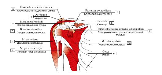 Рис. 249. Синовиальные сумки плечевой области:
1 - Pectoralis major; 2 - Deltoid; 3 - Subdeltoid bursa; 4 - Subacromial bursa; 5 - Acromion; 6 - Subcutaneous acromial bursa; 7 - Coracoid process; 8 - Clavicle; 9 - Subtendinous bursa of subscapularis; 10 - Subscapularis; 11 - Rib
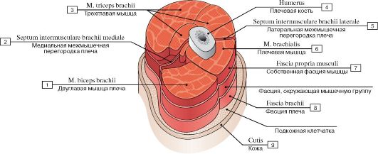 Рис. 250. Фасция мышцы:
1 - Biceps brachii; 2 - Medial intermuscular septum of arm; 3 - Triceps brachii; 4 - Humerus; 5 - Lateral intermuscular septum of arm; 6 - Brachialis; 7 - Fascia of individual muscle; Muscle sheath; 8 - Brachial fascia; 9 - Skin
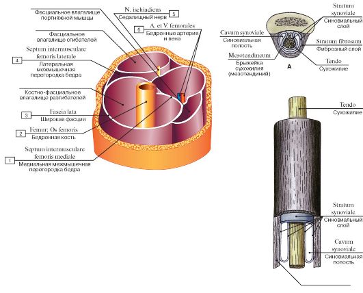 Рис. 251. Костно-фасциальные и фасциальные влагалища мышц нижней трети правого бедра:
1 - Medial femoral intermuscular septum; 2 - Femur; Thigh bone; 3 - Fascia lata; 4 - Lateral femoral intermuscular septum; 5 - Sciatic nerve; 6 - Femoral artery and vein
Stratum fibrosum Фиброзный слой
Рис 252. Синовиальное влагалище сухожилия (А - поперечный разрез, Б - продольный разрез)
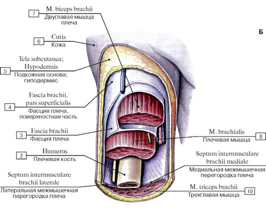 Рис. 253. Поверхностная и глубокая фасции плеча, вид спереди:
1 - Lateral intermuscular septum of arm; 2 - Humerus; 3 - Brachial fascia; 4 - Brachial fascia, superficial part; 5 - Subcutaneous tissue; 6 - Skin; 7 - Biceps brachii; 8 - Brachialis; 9 - Medial intermuscular septum of arm; 10 - Triceps brachii
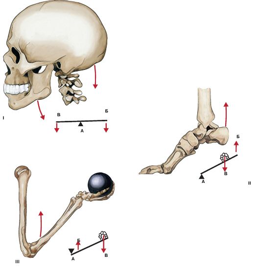 Рис. 254. Схема действия мышц на костные рычаги:
I - рычаг равновесия; II - рычаг силы; III - рычаг скорости; А - точка опоры; Б - точка приложения силы; В - точка
сопротивления
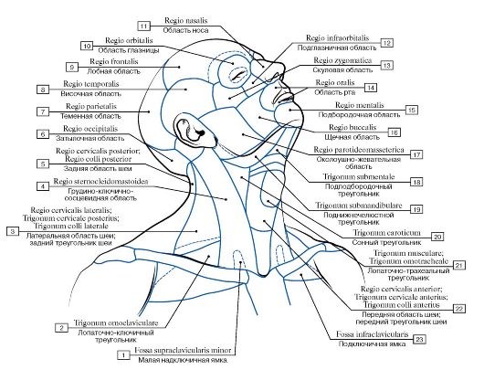 Рис. 255. Области головы и шеи:
1 - Lesser supraclavicular fossa; 2 - Omoclavicular triangle; Subclavian triangle; 3 - Lateral cervical region; Posterior triangle; 4 - Sternocleidomastoid region; 5 - Posterior cervical region; 6 - Occipital region; 7 - Parietal region; 8 - Temporal region; 9 - Frontal region; 10 - Orbital region; 11 - Nasal region; 12 - Infra-orbital region; 13 - Zygomatic region; 14 - Oral region; 15 - Mental region; 16 - Buccal region; 17 - Parotid region; 18 - Submental triangle; 19 - Submandibular triangle; 20 - Carotid triangle; 21 - Muscular triangle; Omotracheal triangle; 22 - Anterior cervical region; Anterior triangle; 23 - Infraclavicular fossa
ив
Рис. 256. Мышцы лица, вид спереди:
1 - Depressor labii inferioris; 2 - Platysma; 3 - Depressor anguli oris; 4 - Risorius; 5 - Levator anguli oris; 6 - Zygomaticus major; 7 - Zygomaticus minor; 8 - Levator labii superioris; 9 - Nasalis; 10 - Levator labii superioris alaeque nasi; 11 - Procerus; 12 - Epicranial aponeurosis; 13 - Occipitofrontalis, frontal belly; 14 - Corrugator supercilii; 15 - Orbicularis oculi; 16 - Buccinator; 17 - Masseter;
18 - Orbicularis oris; 19 - Mentalis
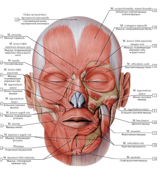
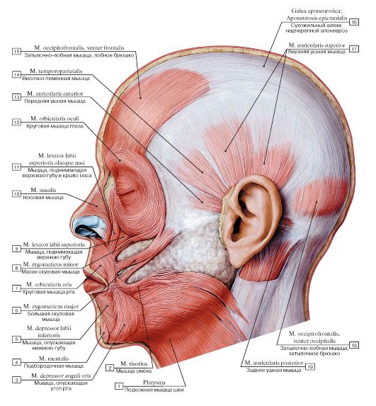 Рис. 257. Мышцы лица, вид слева:
1 - Platysma; 2 - Risorius; 3 - Depressor anguli oris; 4 - Mentalis; 5 - Depressor labii inferioris; 6 - Zygomaticus major; 7 - Orbicularis oris; 8 - Zygomaticus minor; 9 - Levator labii superioris; 10 - Nasalis; 11 - Levator labii superioris alaeque nasi; 12 - Orbicularis oculi; 13 - Auricularis anterior; 14 - Temporoparietalis; 15 - Occipitofrontalis, frontal belly; 16 - Epicranial aponeurosis; 17 - Auricularis superior; 18 - Occipitofrontalis, occipital belly; 19 - Auricularis posterior
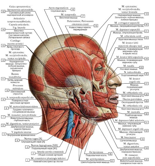 Рис. 258. Мышцы головы, вид справа. На разрезе показаны части жевательной мышцы:
I - Inferior constrictor; 2 - Common carotid artery; 3 - Hypoglossal nerve [XII]; 4 - Vagus nerve [X]; 5 - Internal jugular vein; 6 - Digastric, posterior belly; 7 - Masseter, deep part; 8 - Sternocleidomastoid; 9 - Superficial temporal artery; 10 - Styloid process;
II - Ramus of mandible; 12 - Epicranius; Occipitofrontalis, occipital belly; 13 - Cartilage of acoustic meatus; 14 - Temporomandibular joint; Joint capsule; Articular capsule; Lateral ligament; 15 - Epicranial aponeurosis; 16 - Zygomatic arch; 17 - Temporalis; Temporal muscle; 18 - Pericranium; 19 - Epicranius; Occipitofrontalis, frontal belly; 20 - Corrugator supercilii; 21 - Depressor supercilii; 22 - Orbicularis oculi; 23 - Levator labii superioris alaeque nasi; 24 - Levator labii superioris; 25 - Nasalis; 26 - Infra-orbital nerve; 27 - Levator anguli oris; 28 - Orbicularis oris; 29 - Parotid duct; 30 - Mentalis; 31 - Depressor labii inferioris; 32 - Depressor anguli oris;
33 - Buccinator; 34 - Digastric, anterior belly; 35 - Masseter, superficial part; 36 - Hyoid bone; 37 - Stylohyoid
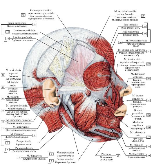 Рис. 259. Мышцы головы, вид справа:
1 - Anterior belly; 2 - Posterior belly; 3 = 1 + 2 - Digastric; 4 - Superficial part; 5 - Deep part; 6 = 4 +5 - Masseter; 7 - Antitragicus; 8 - Auricularis posterior; 9 - Occipitofrontalis, occipital belly; 10 - Tragicus; 11 - Helicic major; 12 - Auricularis superior; 13 - Deep layer; 14 - Superficial layer; 15 = 13 + 14 - Temporal fascia; 16 - Epicranial aponeurosis; 17 - Occipitofrontalis, frontal belly; 18 - Orbital part; 19 - Palpebral part; 20 = 18 + 19 - Orbicularis oculi; 21 - Levator labii superioris; 22 - Levator labii superioris alaeque nasi; 23 - Depressor septi nasi; 24 - Levator anguli oris; 25 - Parotid duct; 26 - Modiolus; 27 - Orbicularis oris; 28 - Mentalis; 29 - Buccinator; 30 - Platysma
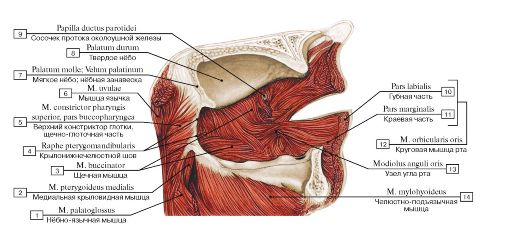 Рис. 260. Мышцы лица со стороны ротовой полости, левая сторона:
1 - Palatoglossus; 2 - Medial pterygoid; 3 - Buccinator; 4 - Pterygomandibular raphe; 5 - Superior constrictor, buccopharyngeal part; 6 - Musculus uvulae; 7 - Soft palate; 8 - Hard palate; 9 - Papilla of parotid duct; 10 - Labial part; 11 - Marginal part; 12 = 10 + 11 - Orbicularis oris; 13 - Modiolus; 14 - Mylohyoid
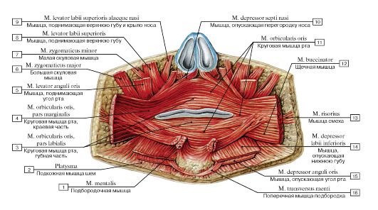 Рис. 261. Мышцы лица, окружающие ротовую щель, вид спереди:
1 - Mentalis; 2 - Platysma; 3 - Orbicularis oris, labial part; 4 - Orbicularis oris, labial part; 5 - Levator anguli oris; 6 - Zygomaticus major; 7 - Zygomaticus minor; 8 - Levator labii superioris; 9 - Levator labii superioris alaeque nasi; 10 - Depressor septi nasi; 11 - Orbicularis oris; 12 - Buccinator; 13 - Risorius; 14 - Depressor labii inferioris; 15 - Depressor anguli oris; 16 - Transversus menti
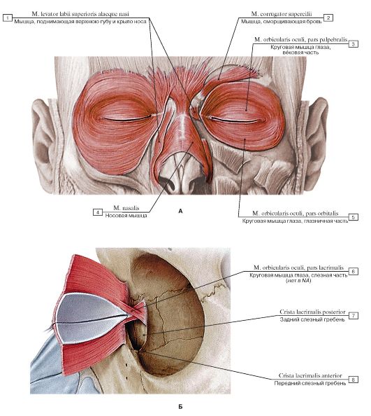 Рис. 262. Мышцы лица, окружающие глазную щель, мышцы окружности ноздрей (А - вид спереди, Б - вид слева):
1 - Levator labi i superioris alaeque nasi; 2 - Corrugator supercilii; 3 - Orbicularis oculi, palpebral part; 4 - Nasalis; 5 - Orbicularis oculi, orbital part; 6 - Orbicularis oculi, lacrimal part; 7 - Posterior lacrimal crest; 8 - Anterior lacrimal crest
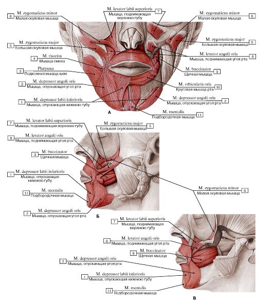 Рис. 263. Мышцы лица, окружающие ротовую щель (А - вид спереди, Б - вид слева, поверхностный слой,
В - вид слева, глубокий слой):
1 - Depressor labii inferioris; 2 - Depressor anguli oris; 3 - Platysma; 4 - Risorius; 5 - Zygomaticus major; 6 - Zygomaticus minor; 7 - Levator labii superioris; 8 - Levator anguli oris; 9 - Buccinator; 10 - Orbicularis oris; 11 - Mentalis
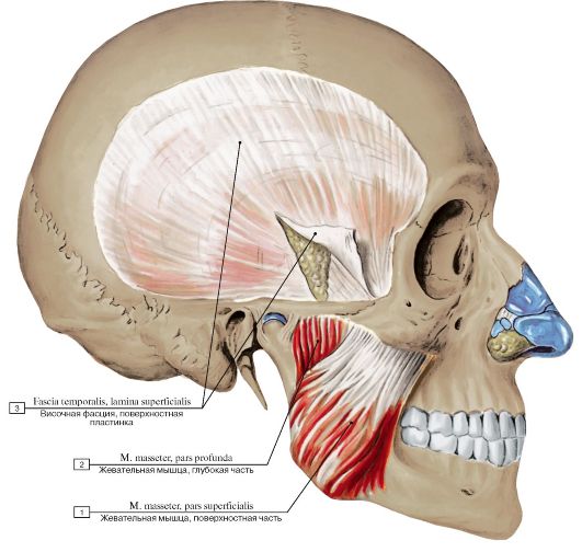 Рис. 264. Жевательные мышцы, вид справа (поверхностный листок височной фасции разрезан и отвернут в сторону):
1 - Masseter, superficial part; 2 - Masseter, deep part; 3 - Temporal fascia, superficial layer
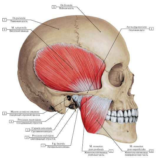 Рис. 265. Жевательная и височная мышцы, вид справа:
1 - Lateral ligament; 2 - Styloid process; 3 - Joint capsule; Articular capsule; 4 - Mastoid process; 5 - External acoustic meatus; 6 - Temporalis; Temporal muscle; 7 - Parietal bone; 8 - Frontal bone; 9 - Zygomatic arch; 10 - Masseter, superficial part; 11 - Masseter, deep
part
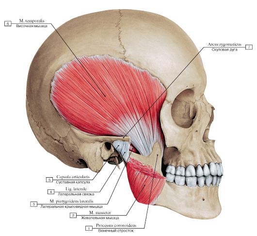 Рис. 266. Височная мышца, вид справа, скуловая дуга отпилена, жевательная мышца частично удалена:
1 - Coronoid process; 2 - Masseter; 3 - Lateral pterygoid; 4 - Lateral ligament; 5 - Joint capsule; Articular capsule; 6 - Temporalis; Temporal muscle; 7 - Zygomatic arch
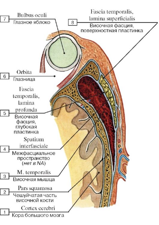 Рис. 267. Поверхностная и глубокая пластинки височной фасции, вид сверху, горизонтальный распил над скуловой дугой:
1 - Cerebral cortex; 2 - Squamous part; 3 - Temporalis; Temporal muscle; 4 - Interfascial space; 5 - Temporal fascia, deep layer; 6 - Orbit; 7 - Eyeball; 8 - Temporal fascia, superficial layer
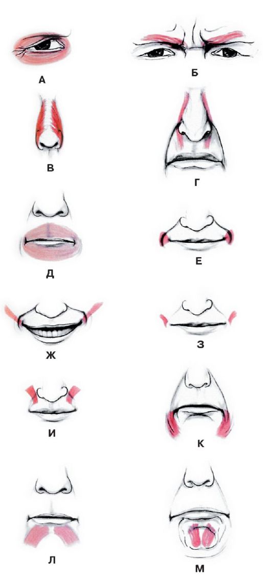 Рис. 268. Изменения выражения лица под действием мышц лица:
А - токращение круговой мышцы глаза в латеральном углу глаза - выражение беспокойства; Б - сокращение мышцы, сморщивающей бровь, происходит в ответ на яркий солнечный свет: «задумчивая бровь»; В - сокращение носовой мышцы вызывает сжатие носа и вызывает радостное или похотливое выражение лица; Г - сильное сокращение мышцы, поднимающей верхнюю губу и крыло носа, с обеих сторон - знак неодобрения; Д - сокращение круговой мышцы рта выражает решительность; Е - сокращение щечных мышц означает чувство удовлетворения; Ж - сокращение большой скуловой мышцы происходит во время улыбки; З - сокращение мышцы смеха отражает целеустремленное действие; И - сокращение мышцы, поднимающей угол рта, выражает самодовольство; К - сокращение мышцы, опускающей угол рта, означает печаль; Л - сокращение мышцы, опускающей нижнюю губу, выражает упорство; М - сокращение подбородочной мышцы означает
упорство
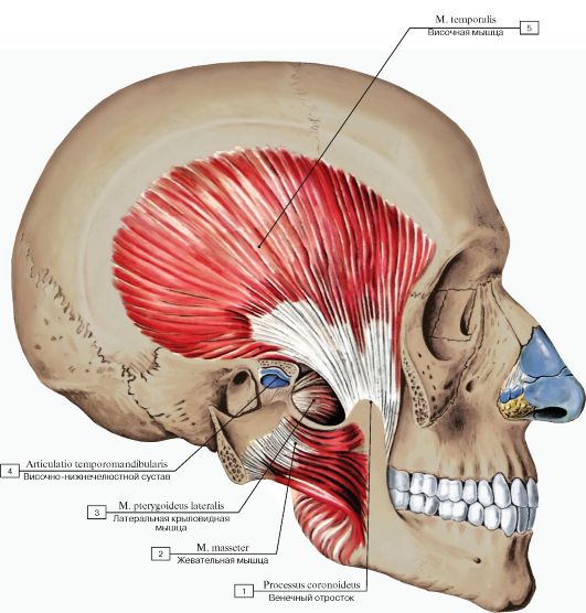 Рис. 269. Жевательные мышцы, вид справа (скуловая дуга отпилена и оттянута в сторону вместе с жевательной мышцей):
1 - Coronoid process; 2 - Masseter; 3 - Lateral pterygoid; 4 - Temporomandibular joint; 5 - Temporalis; Temporal muscle
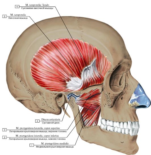 Рис. 270. Жевательные мышцы, вид справа (скуловая дуга и венечный отросток нижней челюсти отпилены и удалены, сухожилие височной мышцы отрезано и поднято кверху):
1 - Medial pterygoid; 2 - Lateral pterygoid, lower head; inferior head; 3 - Lateral pterygoid, upper head; superior head; 4 - Articular disc; 5 - Temporalis; Temporal muscle; 6 - Temporal muscle tendon
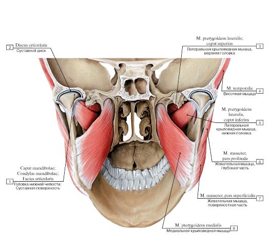 Рис. 271. Жевательные мышцы, вид сзади изнутри:
1 - Head of mandible; Articular surface; 2 - Articular disc; 3 - Lateral pterygoid, upper head; superior head; 4 - Temporalis; Temporal muscle; 5 - Lateral pterygoid, lower head; inferior head; 6 - Masseter, deep part; 7 - Masseter, superficial part; 8 - Medial pterygoid
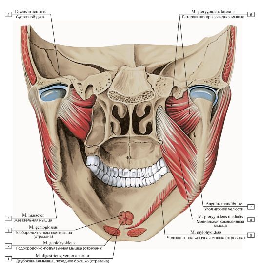 Рис. 272. Жевательные мышцы, вид сзади изнутри:
1 - Digastric, anterior belly; 2 - Geniohyoid; 3 - Genioglossus; 4 - Masseter; 5 - Articular disc; 6 - Lateral pterygoid; 7 - Angle of mandible; 8 - Medial pterygoid; 9 - Mylohyoid
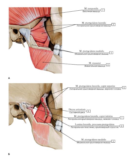 Рис. 273. Латеральная и медиальная крыловидные мышцы, вид справа (А - латеральная и медиальная крыловидные мышцы, Б - медиальная крыловидная мышца):
1 - Temporalis; Temporal muscle; 2 - Lateral pterygoid; 3 - Medial pterygoid; 4 - Masseter; 5 - Lateral pterygoid, upper head; superior head; 6 - Articular disc; 7 - Lateral pterygoid, lower head; inferior head; 8 - Lateral plate, pterygoid process
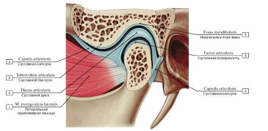 Рис. 274. Прикрепление латеральной крыловидной мышцы к нижней челюсти, вид слева:
1 - Lateral pterygoid; 2 - Articular disc; 3 - Articular tubercle; 4 - Joint capsule; Articular capsule; 5 - Mandibular fossa; 6 - Articular
surface
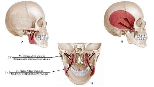 Рис. 275. Схема начала и прикрепления жевательных мышц (А - жевательная мышца, Б - височная мышца, В - латеральные и медиальные крыловидные мышцы):
1 - Medial pterygoid; 2 - Lateral pterygoid
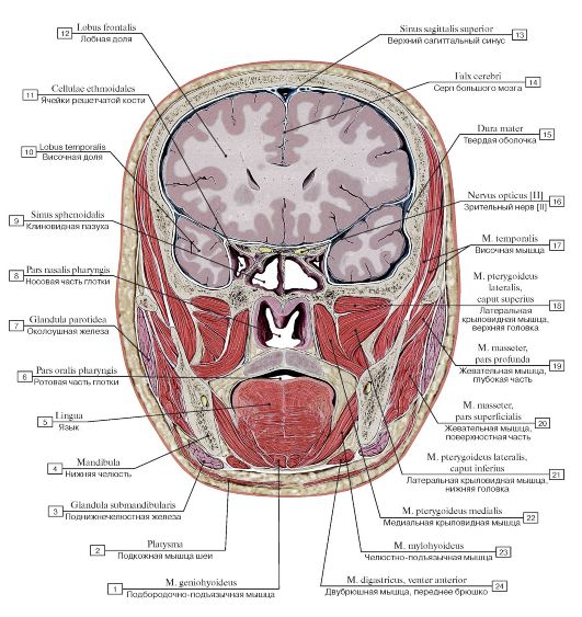 Рис. 276. Фронтальный распил головы на уровне клиновидной пазухи, вид спереди:
1 - Geniohyoid; 2 - Platysma; 3 - Submandibular gland; 4 - Mandible; 5 - Tongue; 6 - Oropharynx; 7 - Parotid gland; 8 - Nasopharynx; 9 - Sphenoidal sinus; 10 - Temporal lobe; 11 - Ethmoidal cells; 12 - Frontal lobe; 13 - Superior sagittal sinus; 14 - Falx cerebri; Cerebral falx; 15 - Dura mater; 16 - Optic nerve [II]; 17 - Temporalis; Temporal muscle; 18 - Lateral pterygoid, upper head; superior head; 19 - Masseter, deep part; 20 - Masseter, superficial part; 21 - Lateral pterygoid, lower head; inferior head; 22 - Medial pterygoid; 23 - My-
lohyoid; 24 - Digastric, anterior belly
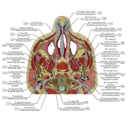 Рис. 277. Горизонтальный распил головы:
1 - Vertebral artery; 2 - Internal jugular vein; 3 - Maxillary vein; 4 - Facial nerve [VII]; 5 - Maxillary artery; 6 - Inferior alveolar artery; 7 - Lingual nerve; 8 - Inferior alveolar nerve; 9 - Masseteric nerve; 10 - Ramus of mandible; 11 - Facial artery; 12 - Facial vein; 13 - Maxillary sinus; 14 - Nasal cavity; 15 - Major alar cartilage; 16 - Nasal septum; 17 - Orbicularis oculi; 18 - Zygomaticus major; 19 - Temporalis; Temporal muscle; 20 - Masseter, deep part; 21 - Masseter, superficial part; 22 - Lateral pterygoid, lower head; inferior head; 23 - Lateral pterygoid, upper head; superior head; 24 - Medial pterygoid; 25 - Parotid gland; 26 - Styloid process; 27 - Vagus
nerve [X]; 28 - Internal carotid artery
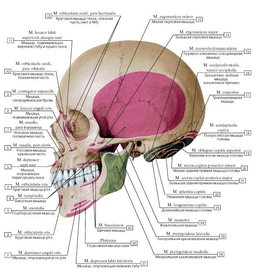 Рис. 278. Места начала и прикрепления мышц на костях черепа, вид сбоку, слева:
1 - Depressor anguli oris; 2 - Orbicularis oris; 3 - Mentalis; 4 - Temporalis; Temporal muscle; 5 - Depressor septi nasi; 6 - Nasalis, alar part; 7 - Nasalis, transverse part; 8 - Levator anguli oris; 9 - Corrugator supercilii; 10 - Orbicularis oculi, orbital part; 11 - Levator labii superioris alaeque nasi; 12 - Orbicularis oculi, lacrimal part; 13 - Zygomaticus minor; 14 - Zygomaticus major; 15 - Sternocleidomastoid; 16 - Occipitofrontalis, occipital belly; 17 - Trapezius; 18 - Semispinalis capitis; 19 - Obliquus capitis superior; 20 - Rectus capitis posterior minor; 21 - Rectus capitis posterior major; 22 - Splenius capitis; 23 - Longissimus capitis; 24 - Masseter; 25 - Buccinator; 26 - Platysma; 27 - Depressor labii inferioris; 28 - Lateral pterygoid; 29 - Medial pterygoid
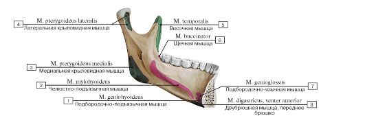 Рис. 279. Места начала и прикрепления мышц на внутренней поверхности нижней челюсти:
1 - Geniohyoid; 2 - Mylohyoid; 3 - Medial pterygoid; 4 - Lateral pterygoid; 5 - Temporalis; Temporal muscle; 6 - Buccinator; 7 - Genioglossus; 8 - Digastric, anterior
belly
 Рис. 280. Наружное основание черепа, вид снизу. Места начала и прикрепления мышц:
1 - Rectus capitis posterior minor; 2 - Semispinalis; 3 - Rectus capitis posterior major; 4 - Obliquus capitis superior; 5 - Splenius; 6 - Longissimus capitis; 7 - Digastric, anterior belly; 8 - Stylohyoid; 9 - Styloglossus; 10 - Temporalis; Temporal muscle; 11 - Lateral pterygoid; 12 - Masseter; 13 - Medial pterygoid; 14 - Longus capitis; 15 - Tensor veli palatini; 16 - Levator veli palatini; 17 - Stylopharyngeus; 18 - Rectus capitis lateralis; 19 - Rectus capitis anterior; 20 - Sternocleidomastoid; 21 - Trapezius
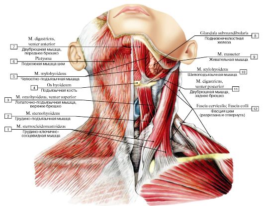 Рис. 281. Подкожная мышца шеи и другие мышцы шеи, вид спереди:
1 - Sternocleidomastoid; 2 - Sternohyoid; 3 - Omohyoid, superior belly; 4 - Hyoid bone; 5 - Mylohyoid; 6 - Platysma; 7 - Digastric, anterior belly; 8 - Submandibular gland; 9 - Masseter; 10 - Stylohyoid; 11 - Digastric, posterior belly; 12 - Cervical fascia
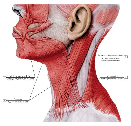 Рис. 282. Подкожная мышца шеи, вид слева:
1 - Platysma; 2 - Depressor anguli oris; 3 - Sternocleidomastoid; 4 - Trapezius
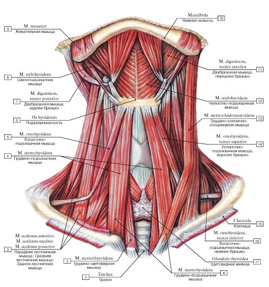 Рис. 283. Поверхностные мышцы шеи, вид спереди. Подкожная и грудино-ключично-сосцевидные мышцы удалены:
I - Trachea; 2 - Sternothyroid; 3 - Scalenus anterior; Anterior scalene; Scalenus medius; Middle scalene; Scalenus posterior; Posterior scalene; 4 - Sternohyoid; 5 - Omohyoid; 6 - Hyoid bone; 7 - Digastric, posterior belly; 8 - Stylohyoid; 9 - Masseter; 10 - Mandible;
II - Digastric, anterior belly; 12 - Mylohyoid; 13 - Sternocleidomastoid; 14 - Omohyoid, superior belly; 15 - Clavicle; 16 - Omohyoid,
inferior belly; 17 - Thyroid gland
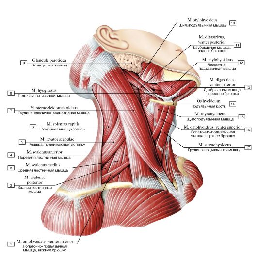 Рис. 284. Грудино-ключично-сосцевидная мышца, надподъязычные и подподъязычные мышцы шеи, вид справа:
I - Omohyoid, inferior belly; 2 - Scalenus posterior; Posterior scalene; 3 - Scalenus medius; Middle scalene; 4 - Scalenus anterior; Anterior scalene; 5 - Levator scapulae; 6 - Splenius capitis; 7 - Sternocleidomastoid; 8 - Hyoglossus; 9 - Parotid gland; 10 - Stylohyoid;
II - Digastric, posterior belly; 12 - Mylohyoid; 13 - Digastric, anterior belly; 14 - Hyoid bone; 15 - Thyrohyoid; 16 - Omohyoid, superior
belly; 17 - Sternohyoid
Рис. 285. Мышцы шеи (мышцы, прикрепляющиеся к подъязычной кости) , вид справа:
1 - Scalenus posterior; Posterior scalene; 2 - Omohyoid, inferior belly; 3 - Levator scapulae; 4 - Longus colli; 5 - Longus capitis; 6 - Semispinalis capitis; 7 - Longissimus capitis; 8 - Splenius capitis; 9 - Sternocleidomastoid; 10 - Digastric, anterior belly; 11 - Stylohyoid; 12 - Masseter; 13 - Inferior constrictor; 14 - Hyoglossus; 15 - Mylohyoid; 16 - Thyrohyoid; 17 - Hyoid bone; 18 - Sternohyoid; 19 - Omohyoid, superior belly; 20 - Sternothyroid; 21 - Thyroid gland; 22 - Tendinous intersection; 23 - Oesophagus; 24 - Trachea; 25 - Clavicle; 26 - First rib [I]; 27 - Scalenus anterior; Anterior scalene; 28 - Scalenus medius; Middle scalene
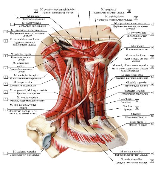
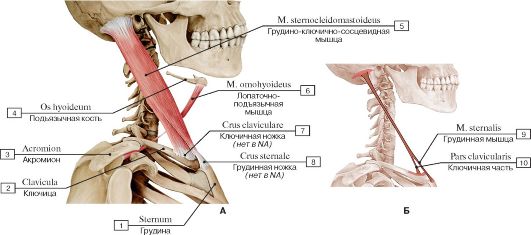 Рис. 286. Грудино-ключично-сосцевидная и лопаточно-подъязычная мышцы, вид справа (А - общий вид, все остальные мышцы удалены, Б - места начала и прикрепления грудино-ключично-сосцевидной мышцы):
1 - Sternum; 2 - Clavicle; 3 - Acromion; 4 - Hyoid bone; 5 - Sternocleidomastoid; 6 - Omohyoid; 7 - Head of clavicle; 8 - Head of
sternum; 9 - Sternalis; 10 - Clavicular head
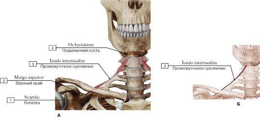 Рис. 287. Лопаточно-подъязычная мышца, вид спереди (А - общий вид, все остальные мышцы удалены, Б - места начала и прикрепления):
1 - Scapula; 2 - Superior border; 3 - Intermediate tendon; 4 - Hyoid bone
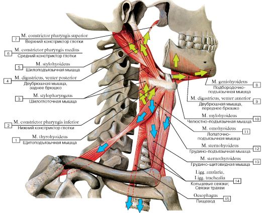 Рис. 288. Направления тяги мышц, прикрепляющихся к подъязычной кости:
1 - Thyrohyoid; 2 - Inferior constrictor; 3 - Stylopharyngeus; 4 - Digastric, posterior belly; 5 - Stylohyoid; 6 - Middle constrictor; 7 - Superior constrictor; 8 - Geniohyoid; 9 - Digastric, anterior belly; 10 - Mylohyoid; 11 - Omohyoid; 12 - Sternohyoid; 13 - Sternothyroid; 14 - Anular ligaments; 15 - Oesophagus
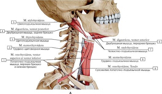
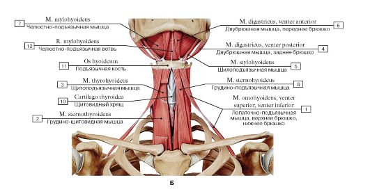 Рис. 289. Над- и подподъязычные мышцы (А - вид справа, Б - вид спереди):
1 - Omohyoid, superior belly, inferior belly; 2 - Sternothyroid; 3 - Thyrohyoid; 4 - Digastric, posterior belly; 5 - Stylohyoid; 6 - Digastric, anterior belly; 7 - Mylohyoid; 8 - Sternohyoid; 9 - Omohyoid; Tendon; 10 - Thyroid cartilage; 11 - Hyoid bone; 12 - Mylohyoid branch
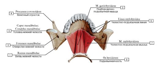 Рис. 290. Челюстно-подъязычные и подбородочно-подъязычные мышцы, вид сверху:
1 - Ramus of mandible; 2 - Mandibular foramen; 3 - Head of mandible; 4 - Coronoid process; 5 - Geniohyoid; 6 - Mylohyoid line;
7 - Mylohyoid; 8 - Hyoid bone
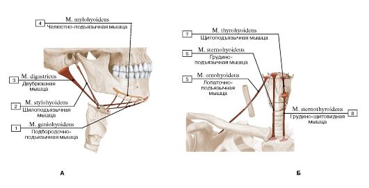 Рис. 291. Места начала и прикрепления (А - надподъязычных мышц, Б - подподъязычн^1х мышц):
1 - Geniohyoid; 2 - Stylohyoid; 3 - Digastric; 4 - Mylohyoid; 5 - Omohyoid; 6 - Sternohyoid; 7 - Thyrohyoid; 8 - Sternothyroid
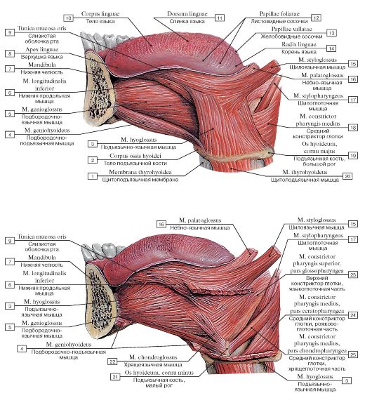 Рис. 292. Дифрагма ротовой полости, вид слева (А - поверхностный слой, Б - глубокий слой):
1 - Thyrohyoid membrane; 2 - Body of hyoid bone; 3 - Hyoglossus; 4 - Geniohyoid; 5 - Genioglossus; 6 - Inferior longitudinal muscle; 7 - Mandible; 8 - Apex of tongue; Tip of tongue; 9 - Mucous membrane of tongue; 10 - Body of tongue; 11 - Dorsum of tongue; 12 - Foliate papillae; 13 - Vallate papillae; 14 - Root of tongue; 15 - Styloglossus; 16 - Palatoglossus; 17 - Stylopharyngeus; 18 - Middle constrictor; 19 - Hyoid bone, greater horn; 20 - Thyrohyoid; 21 - Hyoid bone, lesser horn; 22 - Chondroglossus; 23 - Superior constrictor, glossopharyngeal part; 24 - Middle constrictor, ceratopharyngeal part; 25 - Middle constrictor, chondropharyngeal part
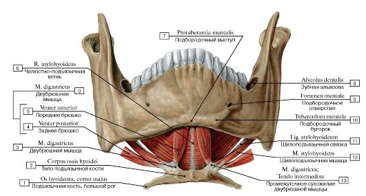 Рис. 293. Надподъязычные мышцы (мышцы диафрагмы ротовой полости), вид спереди и снизу:
1 - Hyoid bone, greater horn; 2 - Body of hyoid bone; 3 - Digastric; 4 - Posterior belly; 5 - Anterior belly; 6 - Mylohyoid branch; 7 - Mental protuberance; 8 - Dental alveolus; 9 - Mental foramen; 10 - Mental tubercle; 11 - Stylohyoid ligament; 12 - Stylohyoid;
13 - Digastric; Intermediate tendon
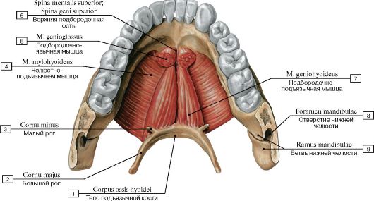 Рис. 294. Надподъязычные мышцы (мышцы диафрагмы ротовой полости), вид сверху:
1 - Body of hyoid bone; 2 - Greater horn; 3 - Lesser horn; 4 - Mylohyoid; 5 - Genioglossus; 6 - Superior mental spine; Superior genial spine; 7 - Geniohyoid; 8 - Mandibular foramen; 9 - Ramus of mandible
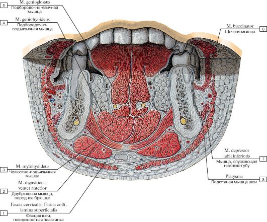 Рис. 295. Мышцы диафрагмы ротовой полости (фронтальный распил через голову на уровне премоляров):
1 - Cervical fascia, investing layer; superficial layer; 2 - Digastric, anterior belly; 3 - Mylohyoid; 4 - Geniohyoid; 5 - Genioglossus; 6 - Buccinator; 7 - Depressor labii inferioris; 8 - Platysma
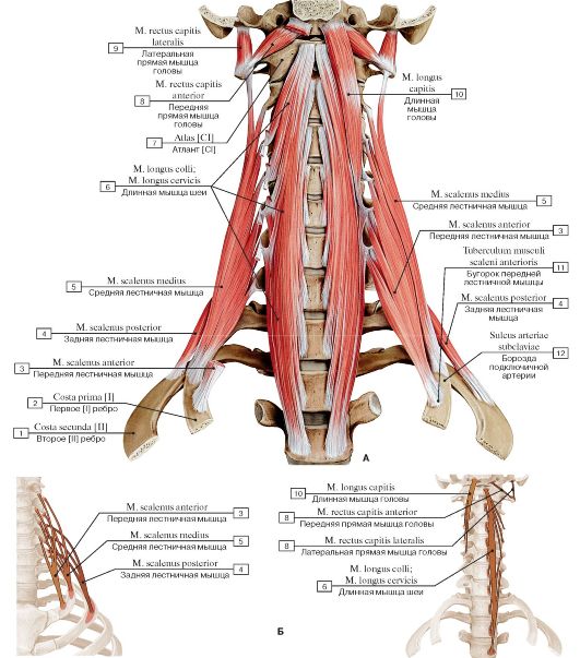 Рис. 296. Глубокие мышцы шеи, вид спереди (А - общий вид, Б - места начала и прикрепления глубоких мышц шеи):
1 - Second rib [II]; 2 - First rib [I]; 3 - Scalenus anterior; Anterior scalene; 4 - Scalenus posterior; Posterior scalene; 5 - Scalenus medius; Middle scalene; 6 - Longus colli; 7 - Atlas [CI]; 8 - Rectus capitis anterior; 9 - Rectus capitis lateralis; 10 - Longus capitis; 11 - Scalene
tubercle; 12 - Groove for subclavian artery
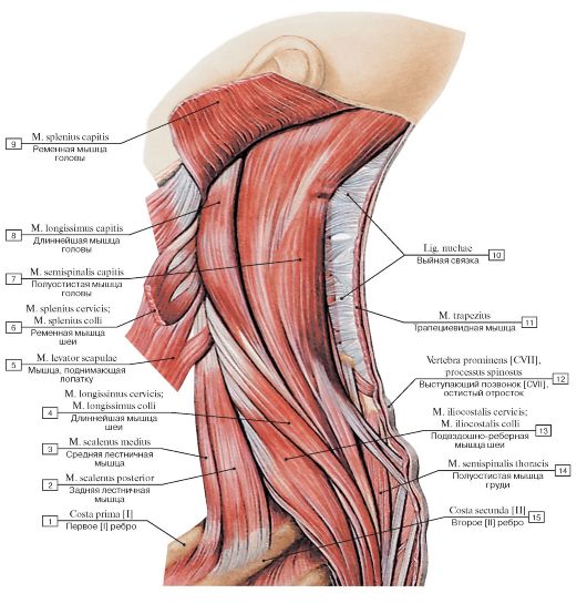 Рис. 297. Мышцы шеи и спины, вид слева:
1 - First rib [I]; 2 - Scalenus posterior; Posterior scalene; 3 - Scalenus medius; Middle scalene; 4 - Longissimus cervicis; 5 - Levator scapulae; 6 - Splenius cervicis; 7 - Semispinalis capitis; 8 - Longissimus capitis; 9 - Splenius capitis; 10 - Ligamentum nuchae; Nuchal ligament; 11 - Trapezius; 12 - Vertebra prominens [CVII], spinous process; 13 - Iliocostalis cervicis; 14 - Semispinalis thoracis; 15 - Second rib [II]
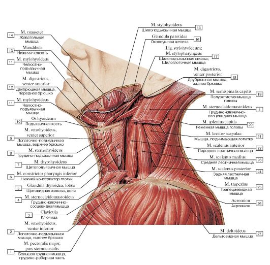 Рис. 298. Глубокие мышцы шеи, вид слева:
1 - Pectoralis major, sternocostal head; 2 - Omohyoid, inferior belly; 3 - Clavicle; 4 - Sternocleidomastoid; 5 - Thyroid gland, lobe; 6 - Inferior constrictor; 7 - Thyrohyoid; 8 - Sternohyoid; 9 - Omohyoid, superior belly; 10 - Hyoid bone; 11 - Mylohyoid; 12 - Digastric, anterior belly; 13 - Mandible; 14 - Masseter; 15 - Stylohyoid; 16 - Parotid gland; 17 - Stylohyoid ligament; Stylopharyngeus; 18 - Digastric, posterior belly; 19 - Semispinalis capitis; 20 - Splenius capitis; 21 - Levator scapulae; 22 - Scalenus anterior; Anterior scalene; 23 - Scalenus medius; Middle scalene; 24 - Scalenus posterior; Posterior scalene; 25 - Trapezius; 26 - Acromion; 27 - Deltoid
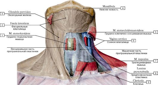 Рис. 299. Пластинки шейной фасции, вид спереди:
1 - Sternohyoid; 2 - Investing layer; 3 - Parotid gland; 4 - Mandible; 5 - Sternocleidomastoid; 6 - Carotid sheath; 7 - Trapezius;
8 - Prevertebral layer; 9 - Clavicle
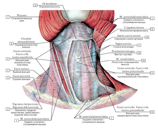 Рис. 300. Фасции шеи, вид спереди:
I - Sternocleidomastoid; 2 - Clavipectoral triangle; Deltopectoral triangle; 3 - Pectoral fascia; 4 - Nuchal fascia; 5 - Cervical fascia, investing layer; superficial layer; 6 - Submandibular gland; 7 - Platysma; 8 - Hyoid bone; 9 - Internal jugular vein; 10 - External carotid artery;
II - Carotid sheath; 12 - Laryngeal prominence; 13 - Omohyoid; 14 - Cervical fascia, pretracheal layer; 15 - Cervical fascia, prevertebral
layer
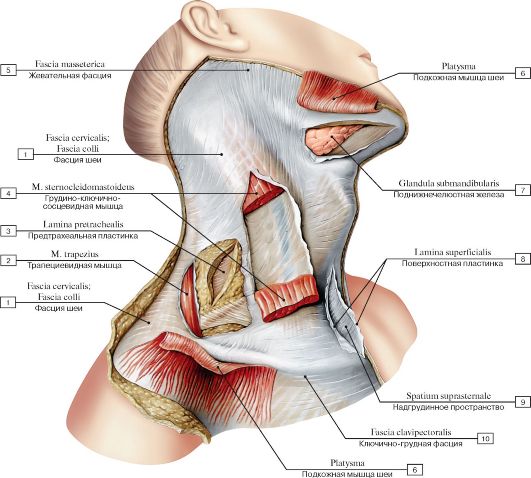 Рис. 301. Фасции шеи, вид спереди и справа. Подкожная мышца шеи и грудино-ключично-сосцевидная мышца отрезаны:
1 - Cervical fascia; 2 - Trapezius; 3 - Pretracheal layer; 4 - Sternocleidomastoid; 5 - Masseteric fascia; 6 - Platysma; 7 - Submandibular gland; 8 - Investing layer; Superficial layer; 9 - Suprasternal space; 10 - Clavipectoral fascia
 Рис. 302. Фасции шеи, вид спереди и справа:
1 - Sternocleidomastoid; 2 - External jugular vein; 3 - Omoclavicular triangle; Subclavian triangle; 4 - Omohyoid, inferior belly; 5 - Cervical fascia, investing layer; superficial layer; 6 - Omohyoid; 7 - Stylohyoid; 8 - Stylohyoid ligament; 9 - Digastric, posterior belly; 10 - Styloglossus; 11 - Styloid process; 12 - Ramus of mandible; 13 - Masseteric fascia; Masseter; 14 - Platysma; 15 - Stylohyoid; Tendon; 16 - Cervical fascia; 17 - Mandible; 18 - Mylohyoid; 19 - Digastric, anterior belly; 20 - Omohyoid, superior belly; 21 - Sternohyoid; 22 - Cervical fascia, p retracheal layer; 23 - Clavicle; 24 - Trachea; 25 - Lesser supraclavicular fossa
Дата добавления: 2017-01-13; просмотров: 11679;