Chromosome mutations and gene mutations
Alterations in the number or structure of chromosomes are called chromosome mutations.Chromosome mutations can happen during mitosis and meiosis when chromosomes are being condensed and pulled apart. Homologous chromosomes may fail to separate, resulting in non-disjunction. Chromosome mutations also occur during interphase when DNA replicates, and during crossing over when sections of chromosomes are exchanged.
Gene mutationsare changes in the nucleotide base sequence in a cistron (the portion of DNA that makes up a single gene). A change of a single nucleotide base pair is called a point mutation.There are a number of types of point mutation, including:
▪ substitution- the replacement of one nucleotide with another containing a different base
▪ deletion - the loss of a nucleotide
▪ insertionor addition- addition of an extra nucleotide.
Sickle-cell anaemia is an example of an inherited condition that results from a substitution. Gene mutations may also result from duplication(repetition of a portion of a nucleotide sequence within a cistron) and inversion(reversal of the portion of the nucleotide sequence in the cistron).
Most mutations, if expressed, are harmful. Note, however, that in diploid organisms such as ourselves, mutations usually result in recessive alleles. These are expressed only in the homozygous condition unless the mutation is on the X chromosome. Many mutations result in a change in the shape of a protein so that the protein cannot function properly (for example, the mutation that causes sickle-cell anaemia). Mutations that affect large sections of a gene, and chromosome mutations are often lethal. However, some mutations have no effect: a mutation may occur in a non-coding part of DNA; it may produce a different codon for the same amino acid; or the altered amino acid sequence may not affect the protein's shape or function. Occasionally, a mutation is beneficial, changing the phenotype so that an organism has a better chance of surviving and reproducing. Although beneficial mutations are very rare events, they are bound to happen sooner or later if there is a large number of individuals in a population. These mutations are of immense importance because they are the ultimate source of all variation: the raw material for the evolution of new species by natural selection.
Down's Syndrome And Genetic Screening
Down's syndrome: trisomy 21
 Down's syndrome is the most common single cause of learning disability in children of school age. Children with the syndrome typically have a round, flat face, and eyelids that appear to slant upwards. In addition to some learning disability, they also have an increased risk of infection (particularly respiratory and ear infections), and heart defects occur in about one-quarter of those with the syndrome.
The syndrome is named after John Langdon Down, a nineteenth century doctor who first described the condition in 1866. In 1959, the French physician Lejeune used chromosome-staining techniques to show that Down's syndrome is caused by an extra chromosome 21. Having one extra chromosome is known as trisomy,hence Down's syndrome is also known as trisomy 21.The extra chromosome usually comes from the egg cell due to non-disjunction of chromosome 21. About 70% of the non-disjunctions occur during meiosis I, when homologous chromosomes fail to separate; 30% occur during meiosis II, when sister chromatids fail to separate. Whether it occurs during meiosis I or meiosis II, non-disjunction leads to trisomy. In a few cases, the extra chromosome comes the father.
In about 3% of cases, Down's syndrome results from translocationof an extra chromosome 21. A region of the chromosome breaks off and rejoins with either the end of the other chromosome 21 or with another non-homologous chromosome (commonly chromosome 15). In these cases, a person may have the normal number of chromosomes, but one of the chromosomes will be abnormally long.
Genetic screening: amniocentesis and chorionic villus sampling
Because of the high risk of Down's syndrome among the babies of older mothers, in the UK mothers over the age of 35 years are usually offered free genetic screening by the National Health Service. Genetic screeningrefers to procedures used to examine an individual for the presence of a genetic disease or disorder. The most widely available genetic screening procedure for Down's syndrome is amniocentesis.
Amniocentesisis usually carried out at 15-16 weeks of pregnancy. It involves passing a very fine needle into the uterus, observed with an ultrasound image, and withdrawing a sample of amniotic fluid containing fetal cells. The karyotype of the fetal cells is then analysed to test for Down's syndrome. The fetal cells can also be cultured in a suitable medium in a laboratory so that further tests, such as DNA analysis, can be carried out.
Amniocentesis is performed under local anaesthetic and most women do not find it too uncomfortable. However, there is a 0.5-1 per cent risk of spontaneous miscarriage after the procedure. Therefore, amniocentesis is usually recommended only for those at high risk of carrying a Down's baby-In the 1970s, chorionic villus sampling (CVS)was developed in China. In CVS, a sample of cells is taken from the chorionic villus (small finger-like processes which grow from the embryo into the mothers uterus). The sample is obtained either by inserting a needle through the abdomen, or inserting a catheter. The fetal cells in the sample can then be analysed in the same way as for amniocentesis.
CVS can be carried out between week 8 and week 12 of pregnancy. If the test shows the fetus has Down's syndrome, a decision about abortion can be made earlier than with amniocentesis. Early abortions are usually less difficult, both physically and mentally, than later abortions. However, a higher risk of miscarriage is associated with CVS than with amniocentesis.
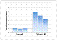
Until recently, a mother's age was the only factor available to assess the risk for Down's syndrome. Now biochemical markers are being discovered for the condition. For example, women with a high risk of Down's syndrome pregnancies tend to have about twice as much chorionic gonadotrophin (a sex hormone produced in placenta cells) in their blood serum as women with normal pregnancies. Tests for these biochemical markers cannot show the presence of a Down's baby, but they can be used in conjunction with the mother's age to predict the probable risk of having a baby with Down's syndrome. If the risk is high, the mother can then decide whether to have an amniocentesis or CVS.
Дата добавления: 2015-11-04; просмотров: 1282;