Chromosomes form homologous pairs
 If the chromosomes are cut out they can be arranged into matching pairs according to their size and certain other features. These are called homologouspairs. Apart from the sex chromosomes, both chromosomes in a pair normally contain the same genes (for example, for eye or hair colour). However, these may be different forms of the gene (for example, one chromosome carries the form for green eyes, the other for brown eyes).
Human cells each have 46 chromosomes (23 pairs). Other species have different numbers, for example, chimpanzee cells each have 48 (24 pairs) and cabbage plant cells each have 18 (9pairs).
One chromosome in each pair comes from the individual’s mother and the other from the father.
- Cells that have the normal two sets of chromosomes are called diploid.
- Cells that give rise to gametes (eggs and sperm) have only one chromosome of each pair, so they have half the normal number of chromosomes. Such cells are calledhaploid.
- In humans, n =23, so normal diploid cells have 46 chromosomes and the haploid gametes have 23 chromosomes.
Mitosis: two identical daughter cells
In mitosis, the nucleus divides once and produces two identical nuclei. The new daughter cells are genetically identical to the parental cell (unless their DNA has been changed in some way, for example by a mutation). So mitosis doubles the number of cells without changing the genetic information. New cells for growth of a multicellular organism, asexual reproduction, and wound healing, for example, are produced by mitosis.
The cell cycle
The cell cycle is the sequence of events that occurs between one cell division and the next. It consists of three main stages:
1. During interphase,the cell grows, carries out its functions, and replicates its DNA. After the DNA is replicated, new protein becomes attached to it. The chromosome now consists of two strands called sister chromatids which contain identical genetic information.Sister chromatids are joined at some point along their length by a centromere. These become visible under the light microscope only during mitosis. Typically, interphase lasts for about 90 per cent of the cell cycle.
2. Nuclear division takes place during mitosis. The chromatids containing replicated DNA are separated from each other and are redistributed as chromosomes in the nuclei of the two new daughter cells.
3. In cell division(also called cytokinesis) the cytoplasm divides to form two daughter cells.
The duration of the cell cycle varies according to conditions such as temperature and the type of cell. The cell cycle of some plant cells (for example, stamen cells of Tradescantia) takes less than 30 minutes at 45°C, but more than two hours at 10°C. Cells in the growing root tip of an onion divide every 22 hours at 20°C. Some cells such as human nerve cells do not divide at all once they have become specialized.
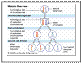
Meiosis: four different daughter cells
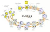 In meiosis, the nucleus divides twice. This produces four haploid nuclei. The number of chromosomes is therefore halved during meiosis. Moreover, homologous chromosomes within a pair can exchange genetic material before being separated. The daughter cells are therefore genetically different from the parent cell (and from each other).
Meiosis is the basis of sexual reproduction, occurring at some point in the life cycle of organisms that reproduce sexually. The haploid gametes produced by meiosis fuse during fertilization. This means that the new fertilized cell has the diploid number of chromosomes. Without meiosis in the life cycle, the number of chromosomes of a sexually reproducing species would be doubled in each generation.
Microscopes
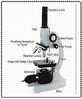 A microscope is used to produce a magnified image of an object or specimen. Anton van Leeuwenhoek (1632-1723) was the first to invent a microscope powerful enough to explore the world of microbes. His discoveries stimulated an explosion of interest in the scientific use of microscopes. Since the 18th century many new types have been invented, of which the most commonly used today are the compound light microscopeand the electron microscope.
Дата добавления: 2015-11-04; просмотров: 1273;