SPINAL CORD
The spinal cord is extremely important to the overall function of the nervous system. It is the communication link between the brain and the peripheral nervous system inferior to the head, integrating incoming information and producing responses through reflex mechanisms.
The spinal cord extends from the foramen magnum to the level of the second lumbar vertebra. It is shorter than the vertebral column because it does not grow as rapidly as the vertebral column during embryonic development. It is composed of cervical, thoracic, lumbar, and sacral segments, which are named according to the area of the vertebral column from which their nerves enter and exit. Because the spinal cord is shorter than the vertebral column, the nerves do not always exit the vertebral column at the same level that they exit the spinal cord. Thirty-one pairs of the spinal nerves exit the spinal cord and pass out of the vertebral column through the intervertebral foramina.
The spinal cord is not uniform in diameter throughout its length. There is a general decrease in diameter superiorly to inferiorly, and there are two enlargements where nerves supplying the limbs enter and leave the cord. The cervical enlargement in the inferior cervical region corresponds to the location at which nerves that supply the upper limbs enter or exit the cord, and the lumbosacral enlargement in the inferior thoracic and superior lumbar regions is the site at which the nerves that supply the lower limbs enter or exit.
Immediately inferior to the lumbar enlargement the spinal cord tapers to form a cone-like region called the conus medullaris. Its tip is at the level of the second lumbar vertebra and is the inferior end of the spinal cord. A connective tissue filament, the filum terminale, extends inferiorly from the apex of the conus medullaris to the coccyx and functions to anchor the cord to the coccyx. The nerves supplying the legs and other inferior structures of the body (L2 to S5) exit the lumbar enlargement and conus medullaris, course inferiorly through the vertebral canal, and exit through the intervertebral foramina from L2 to S5. The conus medullaris and the numerous nerves extending inferiorly from it resemble a horse's tail and are therefore called the cauda equina.
BRAIN
The brain is the part of the CNS located within the cranial vault. The major regions of the adult brain are the cerebrum, the thalamus and hypothalamus, midbrain, pons, medulla oblongata, and cerebellum. The brain works to analyze bits of information before transmitting these messages throughout the body. These messages affect functions such as coordination, learning, memory, emotion, and thought.
The scientists determined the brain was composed of approximately 100 billion neurons, their connections, and supporting cells, which add up to approximately 3 pounds of tissue. This dense network of interconnected neurons is organized to convey all the control signals necessary for individual activities.
The brain is connected to the spinal cord by the brain stem, which is composed of the medulla, the pons, and midbrain. The brain stem controls many of the vital functions, such as breathing and circulation of blood. Cranial nerves exit from the brain stem to control muscles of the face, eyes, tongue, ears, and throat. They also convey sensations from these parts back to the brain.
The cerebrum consists of thick masses of nerve tissue. It is divided into two sides (cerebral hemispheres). Conscious functions such as speech, memory, and vision are controlled in the cerebral hemispheres. Specific areas within these hemispheres are responsible for certain functions, such as speech and the control of muscles in particular parts of the body. In general, control of the muscles of the right side of the body is in the left hemisphere of the brain, and muscles of the left side of the body are controlled by the right hemisphere of the brain. The linking of higher brain functions with cerebral areas is a very active field of research.
The other major portion of the brain, the cerebellum, is located beneath the cerebral hemispheres. It helps control the coordination. At the core of the brain, atop the brain stem, there are other key areas, including thalamus and hypothalamus. The hypothalamus is an endocrine regulatory center that affects sleep and appetite. The thalamus is a collection of nerve cells whose function is the transmission of many of the sensations. In addition, the centers under the cortex play critical roles in relaying messages between different areas of the brain.
Ex. 11. Translate the following words and word-combinations into English:
| Півкуля; ядро; відповідати; середній мозок; кора головного мозку; довгастий мозок; свідомий; зв'язок; поверх, над; передавати, транслювати; склепіння|склепіння,звід| черепа; міст; невелика кількість; мозочок; впливати; головний мозок; центральна нервова система; головні ділянки; набір нервових клітин; передача відчуттів; дослідження; нервова тканина; м’яз; тканина; аналізувати; . | 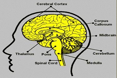 Brain
Brain
|
Дата добавления: 2015-09-11; просмотров: 1214;