DIGESTIVE SYSTEM
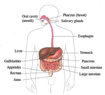
The digestive system consists of many parts. They are the oral cavity, esophagus, stomach, small and large intestines, the liver, the pancreas, gallbladder and others.
The food we eat is propelled through the digestive tract by muscular contractions. The digestive tract is also called the alimentary tract or alimentary canal. The term gastrointestinal tract technically only refers to the stomach and intestines but is often used as a synonym of the digestive tract.
The first division of the digestive tract is the mouth, or oral cavity. Important structures of the oral cavity are the teeth, the tongue, the soft and hard palates, and salivary glands. Digestion begins when the person chews the food. The food is broken into smaller pieces by the teeth and is mixed with saliva secreted by the salivary glands.
From the mouth food passes through the pharynx to the esophagus. The major accessory structures of the pharynx and the esophagus are mucous glands.
The esophagus opens into the stomach. It rests in the upper abdomen. It is a dilated portion of the digestive tract. The stomach receives food from esophagus, and its mixing action reduces the food to a semi-liquid mixture. The stomach walls contain many glands from which acid and enzymes are released into the stomach and mixed with ingested food.
The stomach opens into the small intestine. The small intestine is a thin-walled tube approximately 6.5 meters long. It is located in the lower and central portions of the abdominal and pelvic cavities. It is composed of the duodenum, jejunum, and ileum. The first segment of the small intestine is the duodenum. The major accessory structures in this segment of the digestive tract are the liver, the gallbladder, and the pancreas. The next segment of the small intestine is the jejunum. Small glands exist along its length, and it is the major site of absorption. The last segment of the small intestine is the ileum, which is similar to the jejunum except that fewer digestive enzymes and more mucus are secreted and less absorption occurs in the ileum.
The last section of the digestive tract is the large intestine. It is divided into cecum, colon, and rectum. Its major accessory glands secrete mucus. It absorbs water and salts and concentrates indigested food into feces. The first segment is the cecum, with the attached vermiform appendix. The cecum is followed by colon and rectum. The rectum joints the anal canal, which ends at the anus.
Digestive System
The functions of the digestive system are to ingest food, masticate the food, propel the food through the digestive tract, add secretions to the food and digest the food; and absorb water, electrolytes, and other nutrients from the digested food. Once these useful substances are absorbed, they are transported through the circulatory system to cells where they are used. Undigested matter is moved out of the digestive tract and excreted through the anus. The processes of propulsion, secretion, and absorption are regulated by nervous and hormonal mechanisms.
HEART
The heart is a hollow muscle located in the thoracic cavity between the lungs. The heart is responsible for the circulation of the blood. It is known that the heart is a pump. But it is an extraordinary pump. It weighs only about a pound but the heart of a healthy 70-kg person pumps about 7200 L of blood each day at rate of 5 L per minute. If the heart loses its ability to pump blood for even a few minutes, the life of the individual is in danger.
The heart actually has two pumps. Each pump consists of a pair of chambers formed by muscles. The contraction of these muscles causes the blood to be pumped. The lower chamber is called a ventricle and the upper chamber is called an atrium. The four chambers of the heart are separated by valves. Between the right atrium and the right ventricle there is a one-way valve, called the tricuspid valve. The valve that separates the left atrium from the left ventricle is called the mitral (or bicuspid) valve. The left ventricle is separated from the right ventricle by the interventricular septum.
Venous blood from body flows through the superior vena cava and inferior one into the right atrium, then through the tricuspid valve into the right ventricle. The right ventricle pumps blood through the pulmonary valve, via the pulmonary arteries, into the lungs. From the lungs, blood enters the left atrium via the pulmonary veins and flows through the mitral valve into the left ventricle. The left ventricle pumps oxygen-enriched blood through the aortic valve into the aorta for delivery to the body’s tissues.
The tissue of the heart consists of three layers. The exterior layer is the thin epicardium. The middle layer is the myocardium, the heart muscle itself (from the Greek myo for "muscle" and kardia for "heart"). The inner lining of the heart is the endocardium, a thin, smooth structure. The pericardium is a fibrous sac that surrounds the heart. In the space between the pericardium and the epicardium there is a small amount of fluid.
The heart rate varies depending on activity at any given moment. The control mechanism for the heart rate involves electrical impulses. One of the four chambers of the heart, the right atrium, contains a group of cells called the sinus node. The sinus node produces electrical impulses that signal the muscle of the heart to contract in the pumping cycle. When a person is at rest, the heart pumps more slowly and at a regular rate, about 60 to 80 beats per minute. When a person runs, climbs stairs, or otherwise exert yourself, the sinus node issues electrical “instructions” to increase the pace of the heart in order to provide the muscles and other tissues with the necessary additional blood and its supply of oxygen. The heart rate may increase up to 200 beats per minute if you exert yourself strenuously.
The heart rate may be affected by various factors including tobacco use, caffeine-containing foods, alcohol, and a number of drugs. In addition, the cardiac disorders may produce heart rate problems.
Дата добавления: 2015-09-02; просмотров: 2750;